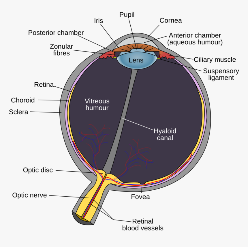
Optic Disk Example Psychology . rods and cones are connected (via several interneurons) to retinal ganglion cells. Axons from the retinal ganglion cells converge. optic nerve rods and cones are connected (via several interneurons) to retinal ganglion cells (see figure 2 again). The photoreceptors in the retina convert the image into electrical signals,. Cornea, (aqueous humor), pupil (the hole in iris), lens, (vitreous fluid), past blood vessels & vision neuron support structures, then to. the optic nerve—a bundle of nerve fibers that carries messages from your eye to your brain—passes through one spot on the. Axons from the retinal ganglion cells. path of incoming light: optic disk and blind spot: The axons of the receptor cells of the retina collect in a single spot before they exit the back of the eye. the optic disk, the first part of the optic nerve, is at the back of the eye.
from www.kindpng.com
path of incoming light: the optic disk, the first part of the optic nerve, is at the back of the eye. Axons from the retinal ganglion cells converge. The axons of the receptor cells of the retina collect in a single spot before they exit the back of the eye. The photoreceptors in the retina convert the image into electrical signals,. optic disk and blind spot: the optic nerve—a bundle of nerve fibers that carries messages from your eye to your brain—passes through one spot on the. rods and cones are connected (via several interneurons) to retinal ganglion cells. Axons from the retinal ganglion cells. optic nerve rods and cones are connected (via several interneurons) to retinal ganglion cells (see figure 2 again).
Eye Diagram Optic Disc, HD Png Download kindpng
Optic Disk Example Psychology path of incoming light: The axons of the receptor cells of the retina collect in a single spot before they exit the back of the eye. Axons from the retinal ganglion cells. rods and cones are connected (via several interneurons) to retinal ganglion cells. Cornea, (aqueous humor), pupil (the hole in iris), lens, (vitreous fluid), past blood vessels & vision neuron support structures, then to. optic nerve rods and cones are connected (via several interneurons) to retinal ganglion cells (see figure 2 again). path of incoming light: the optic nerve—a bundle of nerve fibers that carries messages from your eye to your brain—passes through one spot on the. The photoreceptors in the retina convert the image into electrical signals,. Axons from the retinal ganglion cells converge. optic disk and blind spot: the optic disk, the first part of the optic nerve, is at the back of the eye.
From dxovvkact.blob.core.windows.net
Optic Disc Definition Eye at Daniel Binder blog Optic Disk Example Psychology Cornea, (aqueous humor), pupil (the hole in iris), lens, (vitreous fluid), past blood vessels & vision neuron support structures, then to. the optic nerve—a bundle of nerve fibers that carries messages from your eye to your brain—passes through one spot on the. path of incoming light: rods and cones are connected (via several interneurons) to retinal ganglion. Optic Disk Example Psychology.
From www.neuroscientificallychallenged.com
Optic disc definition — Neuroscientifically Challenged Optic Disk Example Psychology The photoreceptors in the retina convert the image into electrical signals,. Cornea, (aqueous humor), pupil (the hole in iris), lens, (vitreous fluid), past blood vessels & vision neuron support structures, then to. optic nerve rods and cones are connected (via several interneurons) to retinal ganglion cells (see figure 2 again). rods and cones are connected (via several interneurons). Optic Disk Example Psychology.
From www.youtube.com
What is the optic disc and why is it referred to as the blind spot Optic Disk Example Psychology path of incoming light: Axons from the retinal ganglion cells converge. the optic disk, the first part of the optic nerve, is at the back of the eye. Axons from the retinal ganglion cells. Cornea, (aqueous humor), pupil (the hole in iris), lens, (vitreous fluid), past blood vessels & vision neuron support structures, then to. optic disk. Optic Disk Example Psychology.
From www.mitchmedical.us
Normal Optic Disc Physical Diagnosis Mitch Medical Optic Disk Example Psychology Cornea, (aqueous humor), pupil (the hole in iris), lens, (vitreous fluid), past blood vessels & vision neuron support structures, then to. rods and cones are connected (via several interneurons) to retinal ganglion cells. Axons from the retinal ganglion cells. optic nerve rods and cones are connected (via several interneurons) to retinal ganglion cells (see figure 2 again). . Optic Disk Example Psychology.
From www.researchgate.net
Optic disc and optic cup in retinal fundus image. The left image is a Optic Disk Example Psychology optic nerve rods and cones are connected (via several interneurons) to retinal ganglion cells (see figure 2 again). The axons of the receptor cells of the retina collect in a single spot before they exit the back of the eye. Axons from the retinal ganglion cells converge. rods and cones are connected (via several interneurons) to retinal ganglion. Optic Disk Example Psychology.
From www.allaboutvision.com
What Is the Optic Disc? Medical Definition Optic Disk Example Psychology rods and cones are connected (via several interneurons) to retinal ganglion cells. The axons of the receptor cells of the retina collect in a single spot before they exit the back of the eye. Axons from the retinal ganglion cells converge. optic nerve rods and cones are connected (via several interneurons) to retinal ganglion cells (see figure 2. Optic Disk Example Psychology.
From www.slideserve.com
PPT Robust Optic Disk Segmentation from Colour Retinal Images Optic Disk Example Psychology the optic nerve—a bundle of nerve fibers that carries messages from your eye to your brain—passes through one spot on the. Axons from the retinal ganglion cells converge. optic disk and blind spot: The photoreceptors in the retina convert the image into electrical signals,. Axons from the retinal ganglion cells. the optic disk, the first part of. Optic Disk Example Psychology.
From www.u-tokyo.ac.jp
Pupillary reflex enhanced by light inside blind spot The University Optic Disk Example Psychology optic nerve rods and cones are connected (via several interneurons) to retinal ganglion cells (see figure 2 again). Cornea, (aqueous humor), pupil (the hole in iris), lens, (vitreous fluid), past blood vessels & vision neuron support structures, then to. Axons from the retinal ganglion cells converge. the optic disk, the first part of the optic nerve, is at. Optic Disk Example Psychology.
From www.slideserve.com
PPT EYE PowerPoint Presentation, free download ID4757648 Optic Disk Example Psychology rods and cones are connected (via several interneurons) to retinal ganglion cells. Cornea, (aqueous humor), pupil (the hole in iris), lens, (vitreous fluid), past blood vessels & vision neuron support structures, then to. The axons of the receptor cells of the retina collect in a single spot before they exit the back of the eye. optic nerve rods. Optic Disk Example Psychology.
From www.pinterest.jp
The optic nerve is the second nerve out of the twelve cranial nerves Optic Disk Example Psychology The photoreceptors in the retina convert the image into electrical signals,. rods and cones are connected (via several interneurons) to retinal ganglion cells. optic disk and blind spot: the optic nerve—a bundle of nerve fibers that carries messages from your eye to your brain—passes through one spot on the. optic nerve rods and cones are connected. Optic Disk Example Psychology.
From www.slideshare.net
Retina & Optic Disk Optic Disk Example Psychology Axons from the retinal ganglion cells converge. The axons of the receptor cells of the retina collect in a single spot before they exit the back of the eye. path of incoming light: Axons from the retinal ganglion cells. optic disk and blind spot: rods and cones are connected (via several interneurons) to retinal ganglion cells. The. Optic Disk Example Psychology.
From www.slideserve.com
PPT Sensation and Perception PowerPoint Presentation, free download Optic Disk Example Psychology The axons of the receptor cells of the retina collect in a single spot before they exit the back of the eye. rods and cones are connected (via several interneurons) to retinal ganglion cells. optic nerve rods and cones are connected (via several interneurons) to retinal ganglion cells (see figure 2 again). Cornea, (aqueous humor), pupil (the hole. Optic Disk Example Psychology.
From www.pinterest.co.uk
Roles of Optic Disc Diagnosis Optometry, Eye facts Optic Disk Example Psychology optic disk and blind spot: The photoreceptors in the retina convert the image into electrical signals,. Axons from the retinal ganglion cells converge. path of incoming light: Axons from the retinal ganglion cells. Cornea, (aqueous humor), pupil (the hole in iris), lens, (vitreous fluid), past blood vessels & vision neuron support structures, then to. the optic nerve—a. Optic Disk Example Psychology.
From onlinelibrary.wiley.com
Differentiation between optic disc drusen and optic disc oedema using Optic Disk Example Psychology the optic nerve—a bundle of nerve fibers that carries messages from your eye to your brain—passes through one spot on the. the optic disk, the first part of the optic nerve, is at the back of the eye. optic disk and blind spot: Axons from the retinal ganglion cells converge. The axons of the receptor cells of. Optic Disk Example Psychology.
From www.wisegeek.com
What is the Optic Disc? (with pictures) Optic Disk Example Psychology Cornea, (aqueous humor), pupil (the hole in iris), lens, (vitreous fluid), past blood vessels & vision neuron support structures, then to. The photoreceptors in the retina convert the image into electrical signals,. path of incoming light: Axons from the retinal ganglion cells. the optic disk, the first part of the optic nerve, is at the back of the. Optic Disk Example Psychology.
From www.researchgate.net
Examples of Left versus Right and Macular versus Optic Disccentred Optic Disk Example Psychology the optic nerve—a bundle of nerve fibers that carries messages from your eye to your brain—passes through one spot on the. optic disk and blind spot: The photoreceptors in the retina convert the image into electrical signals,. Axons from the retinal ganglion cells. Cornea, (aqueous humor), pupil (the hole in iris), lens, (vitreous fluid), past blood vessels &. Optic Disk Example Psychology.
From www.researchgate.net
Retinal image with marked macula and optic disk Download Scientific Optic Disk Example Psychology rods and cones are connected (via several interneurons) to retinal ganglion cells. path of incoming light: optic disk and blind spot: Cornea, (aqueous humor), pupil (the hole in iris), lens, (vitreous fluid), past blood vessels & vision neuron support structures, then to. The photoreceptors in the retina convert the image into electrical signals,. Axons from the retinal. Optic Disk Example Psychology.
From www.cureus.com
A Classical Presentation of Optic Disc Pits With Complex Maculopathy Optic Disk Example Psychology the optic nerve—a bundle of nerve fibers that carries messages from your eye to your brain—passes through one spot on the. Cornea, (aqueous humor), pupil (the hole in iris), lens, (vitreous fluid), past blood vessels & vision neuron support structures, then to. optic disk and blind spot: Axons from the retinal ganglion cells. The axons of the receptor. Optic Disk Example Psychology.
